Do 3D medical VLMs truly understand volumetric space — or do they rely on language priors?
We introduce CT-SpatialVQA, a benchmark of 9,077 clinically grounded QA pairs
from 1,601 CT volumes that demands explicit spatial reasoning. Eight state-of-the-art models average
only 34% accuracy, often below random chance, revealing a critical gap between
linguistic fluency and volumetric spatial understanding.
CT-SpatialVQA Benchmark
9,077 spatially grounded QA pairs from 1,601 CT volumes with 95% human consensus
Spatially-Grounded QA Design
LLM-based generation, independent validation, and medical human auditing across 6 spatial dimensions
Comprehensive Evaluation
8 medical VLMs benchmarked with LLM-as-Jury and text metrics, revealing consistent failure modes
Method
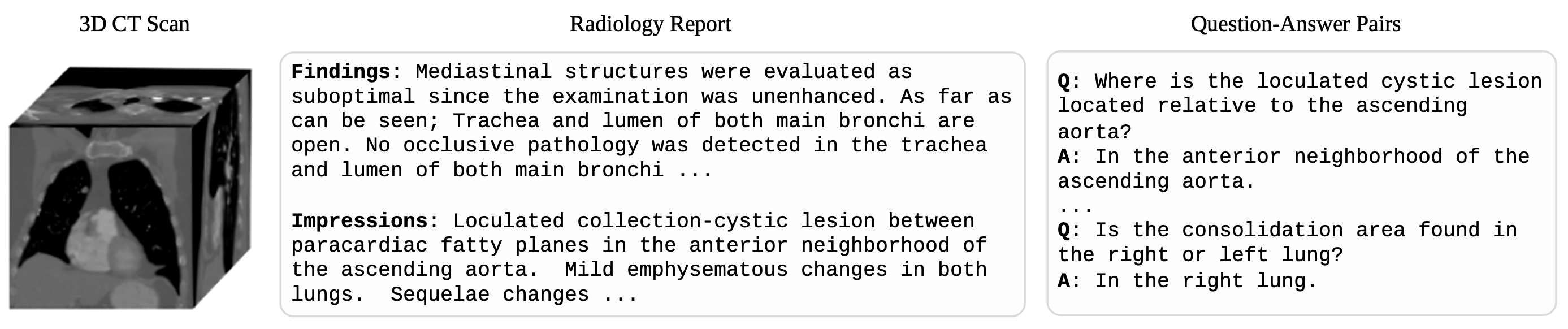
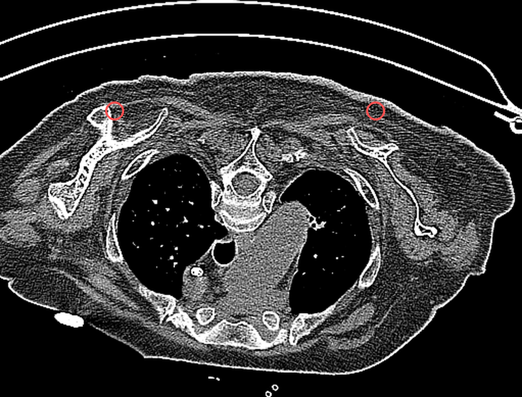
Example. A CT scan, its paired radiology report, and representative spatially-grounded QA pairs.
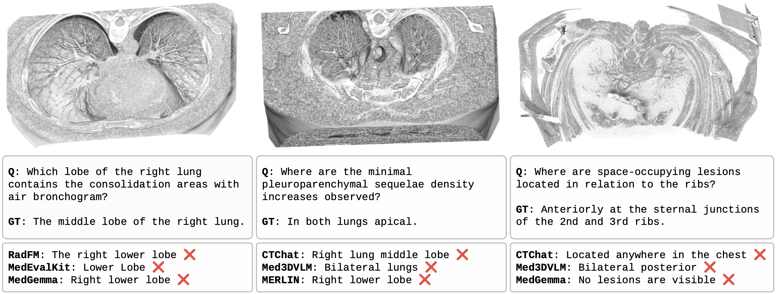
Spatial reasoning failures. All three models answer incorrectly on spatially grounded questions despite varying question types.
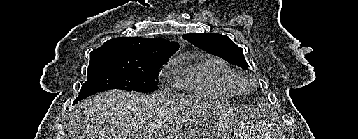
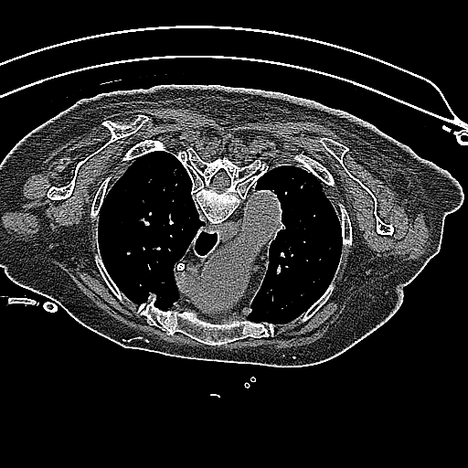
A single CT volume shown across all three anatomical planes:
Axial
Coronal
Sagittal
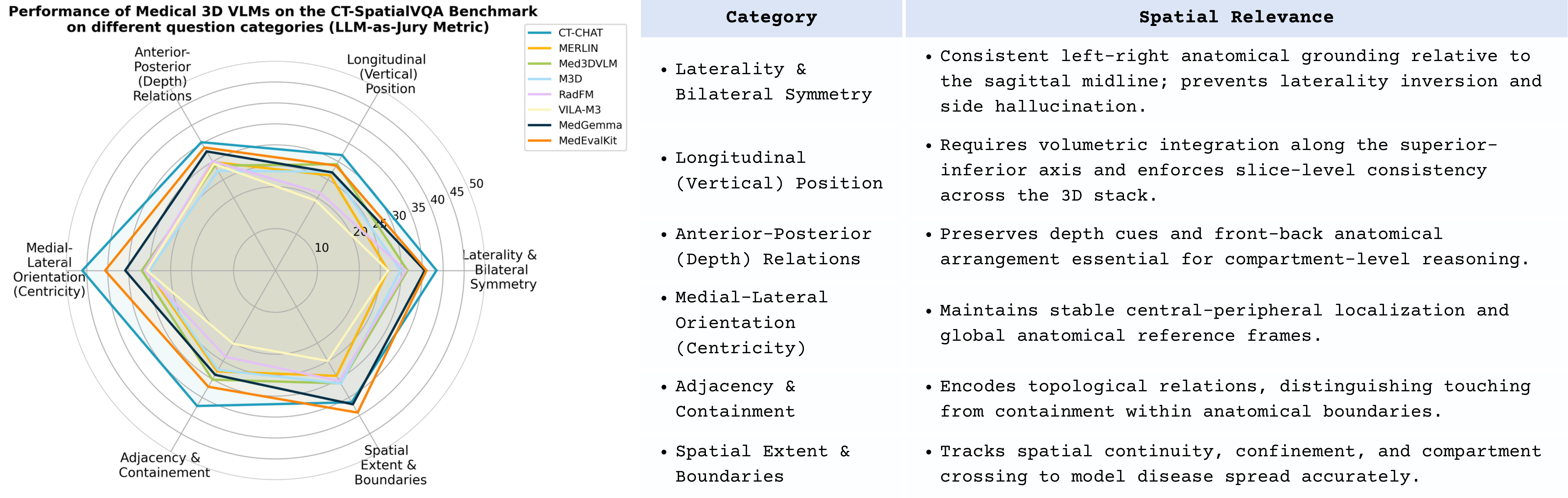
Six Spatial Reasoning Categories
01
Laterality & Bilateral Symmetry
Is the consolidation area in the right or left lung?
In the right lung.
02
Longitudinal (Vertical) Position
Is the pleural effusion located in the upper or lower thorax?
In the lower thorax, predominantly basal.
03
Anterior-Posterior (Depth) Relations
Where is the cystic lesion relative to the ascending aorta?
In the anterior neighborhood of the ascending aorta.
04
Medial-Lateral Orientation (Centricity)
Is the hilar mass centrally or peripherally located?
Centrally located at the hilum.
05
Adjacency & Containment
Where are the space-occupying lesions relative to the ribs?
Anteriorly at the sternal junctions of the 2nd and 3rd ribs.
06
Spatial Extent & Boundaries
Does the ground-glass opacity cross the lobar fissure?
Yes, it extends across the fissure into the adjacent lobe.
Results
Metric
CT-Chat
MERLIN
Med3DVLM
M3D
RadFM
VILA-M3
MedGemma
MedEvalKit
Avg.
LLM-based Evaluation (Accuracy %)
LLM as Judge (Gemini)
44.68
29.85
34.55
33.04
33.36
29.49
39.30
39.96
35.53
LLM as Judge (GPT)
41.50
29.45
31.82
31.09
31.40
28.43
40.55
39.44
34.21
LLM as Judge (Qwen)
45.27
30.34
32.20
31.21
32.09
27.81
37.33
41.89
34.79
LLM as Jury
43.69
28.85
31.44
30.57
31.49
28.20
38.24
40.16
34.08
Text Similarity Metrics
SBERT Cosine Sim.
0.562
0.503
0.399
0.412
0.585
0.471
0.581
0.579
0.512
BLEU
3.29
8.11
0.56
1.08
16.45
2.51
3.07
3.46
4.82
ROUGE-L
0.130
0.300
0.198
0.204
0.372
0.273
0.167
0.158
0.225
METEOR
0.230
0.256
0.068
0.077
0.311
0.114
0.237
0.252
0.193
Category-level performance (LLM-as-Jury). Sharpest drops in vertical and depth-oriented reasoning.
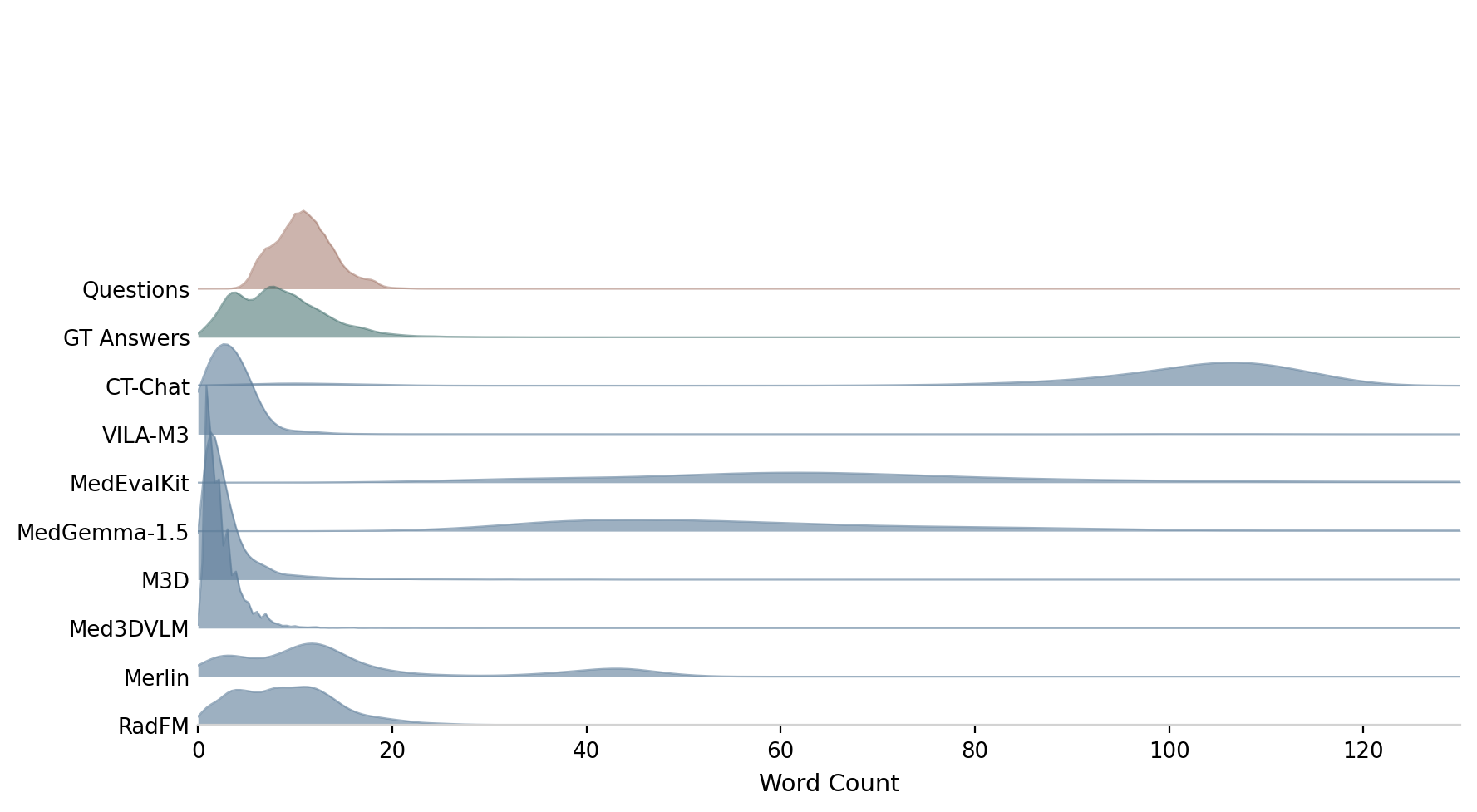
Answer length distribution. Questions, ground-truth answers, and model predictions across all 8 VLMs.
Citation
@article{monon2026ctspatialvqa,
title = {Lost in Volume: The CT-SpatialVQA Benchmark for Evaluating
Semantic-Spatial Understanding of 3D Medical Vision--Language Models},
author = {Monon, Mashrafi and Rahman, Umaima and Hanif, Asif and
Saeed, Numan and Yaqub, Mohammad},
journal = {arXiv preprint arXiv:2605.08787},
year = {2026}
}